


Welcome To McNamara Chiropractic
Stay Active, Stay Strong,
Stay Pain-Free!
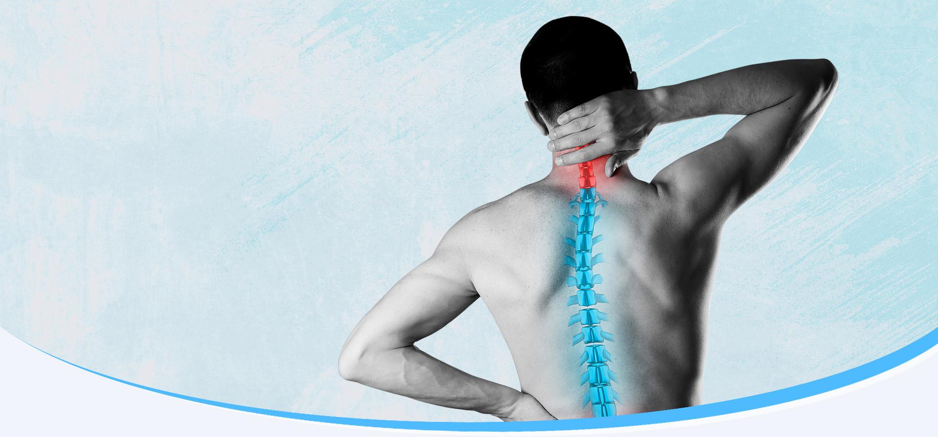
Welcome to McNamara Chiropractic, your trusted source for chiropractic care and Chiropractic Neurology in Cumming, GA. Dr. McNamara is a highly trained Chiropractic Neurologist with expertise in treating everything from simple back pain to complex neurological cases. With hundreds of hours of post-doctorate studies in neurology, concussion and brain trauma rehab, you can trust that you’re in good hands.
Please Call 770-781-4200 To Schedule An Appointment.
more infoWhat we Offer
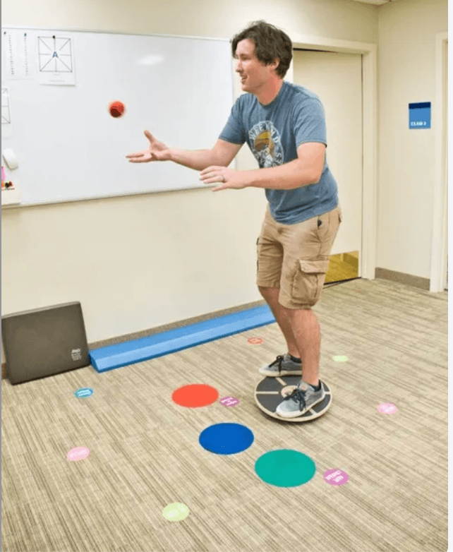
Our Services
Experience the top level of wellness care at McNamara Chiropractic!


Why Choose Us
We help our patients live life pain-free
At McNamara Chiropractic, we understand that overall health is more than just an adjustment. Physical activity, nutrition, and brain health are our main focus. Dr. McNamara is a board-certified Chiropractic Neurologist with hundreds of hours of training in nutritional neurochemistry, movement disorders and post-concussive syndrome.

Quality Care
We believe in quality care tailored to your needs. We listen to our patients and respond with effective treatments that improve their health and well-being.

Informed Patients
We also believe in informing our patients about their health and treatment options. Whether you’re a new patient or experiencing an emergency, we’re always here to help.
Please Call 770-781-4200 To Schedule An Appointment.

Testimonials
What Our Clients Say